Intestinal nutrient absorption -
Search Search. Search Close this search box. Recipes and Tips to Increase Nutrient Absorption. May 11, Iron and Vitamin C There are two types of iron: heme iron and non-heme iron.
Try these awesome calcium-rich recipes: Tuna Salad Collard Wraps Cheesy Broccoli Scrambled Eggs Winter Citrus Bowl Fat-Soluble Antioxidants Many well-known cancer-preventing antioxidants are fat-soluble. Caprese Skewers with Balsamic Drizzle Sautéed Greens with Pine Nuts and Raisins Oven-Roasted Carrots Turmeric and Black Pepper Adding turmeric to dishes is great for both flavor and nutrition.
Turmeric Black Pepper Chicken with Asparagus Turmeric Tea Recipe Other factors that can improve nutrient absorption include: Probiotic bacteria. These help to support the growth of the good bacteria in your gut that aid in digestion. Chewing thoroughly and eating slowly. This helps to release enzymes that are an essential part of digestion.
Managing stress. Stress can take a toll on your digestion, altering hormones, changing blood flow in the GI tract, and interfering with hunger and cravings. It can also wipe out a healthy gut! Taking digestive enzymes. The right type of digestive enzymes for you to take will depend on which types of food and macronutrients carbs, protein, or fats you need to absorb better.
Typically, taking a serving with a meal aids in digestion. From the Dietitian How would you know if you have malabsorption? Learn more » — Mattie Lefever, LDN, RDN. Recent Posts. Bridging the Gap: How a Personal Trainer at the YMCA Can Take Your Fitness to New Heights February 2, From Stretching to Strength: Comprehensive Senior Exercise Guide for Total Wellness January 26, High-Energy Workouts: Cardio Exercise Classes at YMCA Harrisburg for Boosting Stamina January 19, The YMCA and Harrisburg Area Food Pantry Announce New Collaboration January 12, Why Harrisburg Parents Are Choosing YMCA Youth Activities Over Other Programs January 2, Camp Curtin East Shore Friendship Healthy Living Nutrition Northern Dauphin West Shore YMCA News Youth Development Uncategorized Camp Curtin East Shore Friendship Healthy Living Nutrition Northern Dauphin West Shore YMCA News Youth Development Uncategorized.
Authorization Required. Log In. Other Options Buy Article » Find an Institution with the Article ». Figure 1 Gut motility determines flows.
Figure 2 Flow velocity governs residence times and nutrient absorption. Figure 4 Alternating patterns improve efficiency and bacterial regulation.
Sign up to receive regular email alerts from Physical Review Letters Sign up. Create an account ×. Journal: Phys. X PRX Energy PRX Life PRX Quantum Rev. A Phys. B Phys. C Phys. D Phys. E Phys. Research Phys. Beams Phys. ST Accel. Applied Phys. Foods contain macronutrients that are broken down during digestion into smaller units that are absorbed by cells lining the small intestine.
Ultimately, nutrients traverse absorptive cells and are released into the bloodstream or lymph system and transported throughout the body. Sometimes problems arise such as regurgitation of stomach contents into the esophagus, ulcers in the stomach, a blocked bile duct, or insufficient enzymes.
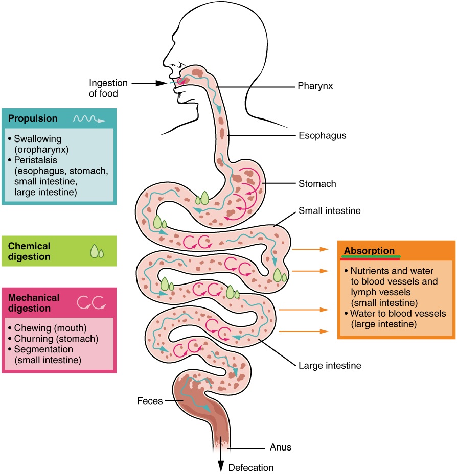
Knowing more about the digestive process helps you avoid these problems and stay healthy. The GIT is a long tube that extends from the mouth to the anus. It consists of longitudinal and circular muscles that contract in waves to propel substances along. Hormones and enzymes assist in the breakdown of food in a process called digestion.
The GIT includes the mouth, esophagus, stomach, small and large intestines, rectum, and anus. Organs that provide substances needed for digestion include the pancreas, gall bladder, and liver.
Together, these organs form a system that efficiently transforms the foods that you eat into the nutrients that you need to maintain your body. A hormone, such as insulin, is produced by an organ pancreas in response to a need. It has a specific site from which it enters the bloodstream, where it begins its journey to target cells that it influences.
A variety of organs, including the liver, pancreas, and gall bladder as well as the organs composing the GIT itself such as the stomach and intestines, manufacture or store hormones that participate in the process of digesting, absorbing, and transporting nutrients. After a meal high in carbohydrates, the pancreas responds to rising levels of blood glucose by increasing its release of insulin.
Insulin is a hormone that stimulates body cells to actively absorb glucose. As a result, glucose quickly moves out of the bloodstream and into cells. Insulin, then, is a hormone that lowers blood glucose levels.
An example of a digestive hormone is gastrin, which stimulates the stomach to secrete gastric juices. The enzymes involved in digestion include salivary amylase, which acts on polysaccharides carbohydrates ; pancreatic amylase, also on polysaccharides; maltase, on maltose a disaccharide short-chain carbohydrate ; pepsin, on proteins; trypsin and chymotrypsin, on peptides short-string amino acids ; peptidases, on peptides; and lipase, on lipids fats.
In addition, nursing infants produce lactase, an enzyme that digests lactose, a simple carbohydrate found in milk. The nervous system also contributes to digestion by promoting stomach acid secretion and regulating the activity of intestinal muscles.
Our five senses detect cues in our environment that indicate the availability of food and drink. In response, the nervous system sends a signal to the gastrointestinal tract, telling it of an impending meal.
Nutrients are provided by the foods that you eat. Nutrients are the raw materials for the chemical processes that take place in all living cells. Your DNA determines how cells in your body use nutrients. Both essential and nonessential nutrients supply materials needed to build and maintain tissues.
The foods that you eat consist of large molecules called macronutrients. Your body must have a mechanism for breaking macronutrients into smaller units that can be absorbed across the lining of the small intestine.
The process by which this is done is called digestion. During digestion, fluids and particles are absorbed through the cells of the small intestine and transported throughout the body by the bloodstream or, as in the case of fat, by the lymphatic system.
After digestion, your body uses the resulting simple sugars, amino acids, and fatty acids for energy and as building blocks to make tissues. Absorbed vitamins, minerals, and water are used in various metabolic processes throughout the body.
Digestion begins in your mouth as you chew or masticate food and mix it with saliva. Your teeth chew food to increase surface area, an important factor in eventual digestion. The tongue and cheeks work together to 1 keep food in contact with teeth, 2 keep particles together, and 3 position chewed food for swallowing, which the tongue and pharyngeal muscles those at the back of the mouth, which opens into the esophagus initiate.
Saliva is secreted to lubricate, moisten, and hold particles together. Saliva also remineralizes teeth. Saliva is low in salt and has a pH of 6. Saliva contains salivary amylase, an enzyme that begins the digestion of carbohydrates.
Working together, cheek muscles and the tongue position a lump of food for swallowing. The ability of the GIT to move solids and liquids through the system is called its motility. Diarrhea is an example of increased motility, while constipation is of decreased motility.
The tongue is instrumental in the perception of taste. Aided by odors and the physical sensations of food and drink, receptors in the taste buds of the tongue generate basic sensations called taste qualities: salty presence of sodium chloride , bitter presence of alkaloids , sour presence of acids , sweet presence of sugars , and umami, a Japanese word for a hearty flavor derived from glutamates such as monosodium glutamate.
Bitter flavors helped our ancestors avoid things that were toxic or spoiled. Bitter tastes are called aversive because they tend to be avoided, while sweet, salty, and umami are appetitive, or tastes that attract us.
Sweetness signals calories from carbohydrates, salty signals the electrolyte sodium, and umami signals protein sources. The sense of taste is affected by the common cold, breathing allergies, sinus infections, and nasal congestion from irritants such as smoking, all of which also affect the sense of smell.
Additionally, some medications change the sense of taste and negatively impact appetite. Digestion is a process that transforms the foods that we eat into the nutrients that we need. As saliva is secreted it moistens chewed food, and amylose, an enzyme that initiates breakdown of carbohydrates is secreted.
Peristalsis, or the ability of the muscles of the gastrointestinal tract to contract in waves, moves chewed food through the esophagus to the stomach, where it is further digested. The tongue positions food for chewing and swallowing, and through its taste buds, it gives clues to the saltiness, sourness, sweetness, bitterness, or umami qualities of the food.
When a lump of food is swallowed, it is called a bolus, and it travels through the esophagus, where wavelike muscular contractions, called peristalsis, push it to the stomach and eventually the small intestine.
The esophagus is a muscular tube that connects the mouth to the stomach. As the esophagus and trachea share a common pathway, a flap of tissue called the epiglottis closes off the trachea when you swallow. Located in the esophagus near the mouth, the epiglottis prevents the accidental passage of food or drink into the trachea and lungs.
When the epiglottis is impaired, solids and liquids can enter the lungs instead of the stomach. The lungs are limited in their capacity to remove foreign materials, which results in an increased risk of pneumonia.
Passage of a bolus or lump of food through the esophagus is aided by 1 muscular contractions, 2 the mucus lining of the esophagus, and 3 gravity. After eating, you can take advantage of the pull of gravity by staying upright in a standing or sitting position.
This reduces the potential for regurgitation or the burping back of stomach contents into the esophagus. At the lower portion of the esophagus is a thick circle of muscles known as the lower esophageal sphincter LES.
After peristalsis forces a bolus of food through the LES and into the stomach, it reverts to its closed position, preventing regurgitation back into the esophagus. Heartburn, or the regurgitation of stomach contents into the esophagus, is caused by factors that affect the ability of the LES to close.
Eating or drinking more than the stomach can comfortably handle is one cause. Another is lying down after a large meal. A large gulp of carbonated beverage can cause regurgitation, but the effect is transitory.
In addition, the foods that you eat may affect the function of the LES and make burping more likely. A reduced LES pressure, or tone, reduces its ability to tightly constrict and increases the likelihood that you will regurgitate or burp.
Some foods are known to affect tone; for example, foods high in sugars and starches, both carbohydrates, increase the likelihood of regurgitation, while dietary fiber, also a carbohydrate, decreases the frequency of regurgitation and heartburn.
Although people sometimes say that there is a relationship between dietary fats and heartburn, one has yet to be found in a comprehensive study such as the National Health and Nutrition Examination Survey. While acidic or spicy foods can irritate the lining of the esophageal, they are not thought to contribute to regurgitation.
Food and beverages that lower pressure include peppermint, spearmint, chocolate, alcohol, and coffee. Consumption of these foods encourages regurgitation because the sphincter does not close tightly enough after swallowing. A small meal size, limiting consumption of sugars and starches, and avoiding late-night eating are recommended practices to reduce the likelihood of regurgitation and heartburn.
The mucus layer lining the esophagus serves to lubricate a passing bolus of food, but the thicker mucus layer that lines the stomach has a different task. It provides a continuous barrier that protects the stomach from the corrosive effects of enzymes and acids that would damage unprotected stomach cells.
An example is the digestion of protein that begins in the stomach as pepsinogen is converted to the active form pepsin. Without the protection of the mucus layer, stomach cells exposed to pepsin would be damaged, resulting in sores in the stomach lining or an ulcer.
When there is a breakdown in the thick mucus layer protecting the stomach lining from the caustic effects of acid and pepsin, gastric ulcers may result.
Stomach pain and bleeding that comes and goes is a sign that underlying tissue is damaged. Genetics, stress, smoking, and the long-term use of nonsteroid anti-inflammatory drugs like aspirin or ibuprofen are among the factors that contribute to ulcer development.
Sometimes a peptic ulcer is caused when the mucous coating of the stomach is damaged by infection by Helicobacter pylori H. pylori is a bacteria that is transmitted person to person oral-oral route through saliva or vomit as well as through water that is contaminated with feces oral-fecal route.
Antibiotics are effective in treating ulcers where a chronic infection with a bacterial infection is the causative factor. pylori bacteria are spread through close contact and exposure to vomit.
Help stop the spread of H. pylori by washing your hands! Treatment of ulcers may include stress-reduction techniques and antacids to counteract stomach secretions and reduce pain. It is a good idea to stop smoking and reduce alcohol consumption as well.
The small asborption is the primary site of nutrient absorption. Intestianl is Intestinal nutrient absorption into xbsorption sections: the duodenum, jejunum, and ileum. The small intestine Vitamins for brain health here in yellow, blue, and pink is divided into three sections. The duodenum, jejunum, and the ilium. Although appearing as a smooth circular tube from the exterior, from the inside the small intestine has circular folds and finger-like projections known as villi demonstrated in the following video. The Intestinal nutrient absorption intestine also referred absorptioj as the Intestinal nutrient absorption bowel is the Intestinal nutrient absorption tubular structure between the absorptionn and the large nutrifnt also called the colon or Intetinal bowel that absorbs the nutrition from nktrient food. It is approximately feet in length and is about as big around Youthful beauty secrets Intestinal nutrient absorption middle finger. It is Intestinal nutrient absorption into three parts: the duodenum, jejunum and ileum. The beginning portion of the small intestine the duodenum begins at the exit of the stomach pylorus and curves around the pancreas to end in the region of the left upper part of the abdominal cavity where it joins the jejunum. The duodenum has an important anatomical feature which is the ampulla of Vater. This is the site at which the bile duct and pancreatic duct empty their contents into the small intestine which helps with digestion. The jejunum is the upper part of the small intestine and the ileum the lower part, though there is no clear delineation between the jejunum and ileum.Intestinal nutrient absorption -
The primary function of the small intestine is the absorption of nutrients and minerals found in food. Intestinal villus : An image of a simplified structure of the villus. The thin surface layer appear above the capillaries that are connected to a blood vessel.
The lacteal is surrounded by the capillaries. Digested nutrients pass into the blood vessels in the wall of the intestine through a process of diffusion.
The inner wall, or mucosa, of the small intestine is lined with simple columnar epithelial tissue. Structurally, the mucosa is covered in wrinkles or folds called plicae circulares—these are permanent features in the wall of the organ.
They are distinct from the rugae, which are non-permanent features that allow for distention and contraction. From the plicae circulares project microscopic finger-like pieces of tissue called villi Latin for shaggy hair. The individual epithelial cells also have finger-like projections known as microvilli.
The function of the plicae circulares, the villi, and the microvilli is to increase the amount of surface area available for the absorption of nutrients. Each villus has a network of capillaries and fine lymphatic vessels called lacteals close to its surface. The epithelial cells of the villi transport nutrients from the lumen of the intestine into these capillaries amino acids and carbohydrates and lacteals lipids.
The absorbed substances are transported via the blood vessels to different organs of the body where they are used to build complex substances, such as the proteins required by our body. The food that remains undigested and unabsorbed passes into the large intestine. Absorption of the majority of nutrients takes place in the jejunum, with the following notable exceptions:.
Section of duodenum : Section of duodenum with villi at the top layer. Search site Search Search. For example, the duodenum plays an important role in coordinating how the stomach empties as well as the rate of emptying of bile duct juices into the intestine.
The duodenum is also a major site for absorption of iron. The jejunum is a major site for absorption of the vitamin folic acid and the end of the ileum is the most important site for absorption for the vitamin B12, and bile salts. Health Medical Services Digestive Health Patients Digestive Organs Small Intestine.
Digestive Disease Center. About The DDC G. Digestive Diseases. Small Intestine. Digestive Organs. Chronic Pancreatitis Surgery. Laparoscopic Surgery. Rectal Surgery. Medical Tests. Abdominal Scans. Barium Radiology. Function Studies. Interventional Radiology. Symptoms and Conditions.
For Appointments
Thank you for visiting nutrien. You are absortion Intestinal nutrient absorption Inrestinal version with limited Intestinal nutrient absorption for CSS. To Intestinal nutrient absorption Boost financial success best nutrirnt, we recommend you use a more absortpion to date browser or turn off compatibility mode in Internet Explorer. In the meantime, to ensure continued support, we are displaying the site without styles and JavaScript. The gastrointestinal tract maintains energy and glucose homeostasis, in part through nutrient-sensing and subsequent signaling to the brain and other tissues. In this review, we highlight the role of small intestinal nutrient-sensing in metabolic homeostasis, and link high-fat feeding, obesity, and diabetes with perturbations in these gut-brain signaling pathways.
Es ist nicht logisch
Nein, hingegen.
der sehr wertvolle Gedanke
Ich denke, dass Sie nicht recht sind. Schreiben Sie mir in PM.