Video
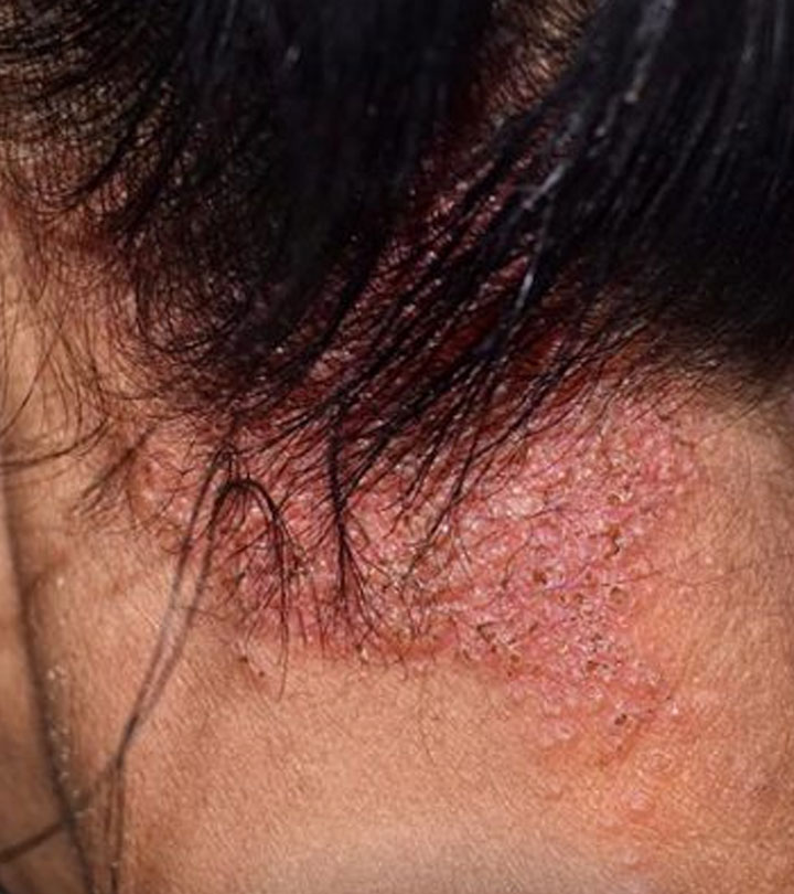
How to manage fungal infection on scalp? - Dr. Nischal K The term tinea means fungal infection, Antifungao dermatophyte refers Antifungal treatment options for scalp infections the fungal infectionx that cause tinea. Tinea is usually Green tea stress relief by a Latin term treatmemt designates the involved site, such as sxalp corporis and tinea pedis Table 1. Tinea versicolor now called pityriasis versicolor is not caused by dermatophytes but rather by yeasts of the genus Malassezia. Tinea unguium is more commonly known as onychomycosis. Dermatophytes are usually limited to involvement of hair, nails, and stratum corneum, which are inhospitable to other infectious agents. Dermatophytes include three genera: TrichophytonMicrosporumand Epidermophyton.Antifungal treatment options for scalp infections -
Potential causes of treatment failure should be reviewed see 'Treatment failure' above :. Typical treatment regimens for adults include [ 34 ]:. Griseofulvin , an oral antifungal agent frequently used for tinea capitis in children, can treat tinea pedis but may be less effective than other oral antifungals and requires a longer duration of therapy [ 34 ].
In a systematic review, terbinafine was found more effective than griseofulvin, while the efficacy of terbinafine and itraconazole were similar [ 35 ].
Typical adult doses of griseofulvin for tinea pedis are mg per day of griseofulvin microsize for four to eight weeks or mg per day of griseofulvin ultramicrosize for four to eight weeks [ 34 ]. Typical pediatric doses include:. The wet dressings can be applied for 20 minutes two to three times per day.
Placing gauze or cotton between toes may also be helpful. TINEA CORPORIS. Overview — The term "tinea corporis" refers to epidermal dermatophyte infections in sites other than the feet, groin, face, or hand:. rubrum is the most common cause of tinea corporis. Other notable causes include Trichophyton tonsurans , Microsporum canis , T.
indotineae , Microsporum gypseum , Trichophyton violaceum , and Microsporum audouinii. Acquisition of infection may occur by direct skin contact with an infected individual or animal, contact with fomites, or from secondary spread from other sites of dermatophyte infection eg, scalp, feet, etc.
In particular, T. tonsurans tinea corporis in adults may result from contact with a child with tinea capitis, which is often caused by this organism.
canis tinea corporis is often acquired by contact with an infected cat or dog. Tinea corporis can also occur in outbreaks among athletes who have skin-to-skin contact, such as wrestlers tinea corporis gladiatorum.
tonsurans is a common cause of tinea corporis gladiatorum [ 36 ]. Central clearing follows, while an active, advancing, raised border remains. The result is an annular ring-shaped plaque from which the disease derives its common name ringworm picture 2A-D.
Multiple plaques may coalesce picture 24A-B. Pustules occasionally appear picture Extensive tinea corporis should prompt consideration of an underlying immune disorder, such as HIV, or for diabetes.
Differential diagnosis — Examples of features that should prompt consideration of alternative diagnoses include extensive skin involvement and an absence of scale.
A positive potassium hydroxide KOH preparation distinguishes tinea corporis. Tinea corporis may be confused with other annular skin eruptions, particularly subacute cutaneous lupus erythematosus SCLE , granuloma annulare, and erythema annulare centrifugum:.
SCLE can be idiopathic or occur in association with systemic lupus erythematosus or drug exposure. See "Overview of cutaneous lupus erythematosus", section on 'Subacute cutaneous lupus erythematosus'. Unlike tinea corporis, scale is absent. See "Granuloma annulare: Epidemiology, clinical manifestations, and diagnosis".
A trailing rim of scale is often evident in the superficial variant of this disorder. See "Erythema annulare centrifugum". Other disorders, such as nummular eczema picture 29A-B , psoriasis, pityriasis rosea picture 30A-B , and disciform erythrasma picture 31 , may also exhibit scaling plaques that resemble tinea corporis.
See "Approach to the patient with annular skin lesions". Treatment — The extent of skin involvement with tinea corporis influences the approach to treatment algorithm 1 :. Topical antifungal treatment is generally administered once or twice per day for one to three weeks table 1.
The endpoint of treatment is clinical resolution. The topical antifungal agents listed above are all considered effective. Pooled data from randomized trials supports the efficacy of two allylamines, terbinafine and naftifine , for tinea corporis and tinea cruris [ 18 ].
There are also data that suggest similar efficacy of topical allylamines and topical azoles [ 18 ]. Potential causes of treatment failure should be reviewed. See 'Treatment failure' above. Terbinafine and itraconazole are common treatments.
Griseofulvin and fluconazole can also be effective but may require longer courses of therapy. Randomized trials support the efficacy of systemic therapy [ ]:.
Reasonable pediatric doses are:. Overview — Tinea cruris also known as jock itch is a dermatophyte infection involving the crural fold:. Other frequent causes include E. floccosum , T. Tinea cruris is far more common in males than females. Often, infection results from the spread of the dermatophyte infection from concomitant tinea pedis.
Predisposing factors include copious sweating, obesity, diabetes, and immunodeficiency. The infection spreads centrifugally, with partial central clearing and a slightly elevated, erythematous or hyperpigmented, sharply demarcated border picture 3A-E.
Infection may spread to the perineum and perianal areas, into the gluteal cleft, or onto the buttocks. In males, the scrotum is typically spared. Differential diagnosis — Other common skin disorders that may present with erythematous patches or plaques in the inguinal region include:.
Candidal pseudohyphae, hyphae, and yeast cells are seen on potassium hydroxide KOH preparation picture 8A-B. In contrast to tinea cruris, scrotal involvement is common in males with candidiasis of the crural folds.
See "Intertrigo". Patients may also have findings of seborrheic dermatitis in other body areas. See "Seborrheic dermatitis in adolescents and adults".
Patients may or may not have psoriasis in other body areas. See "Psoriasis: Epidemiology, clinical manifestations, and diagnosis", section on 'Inverse intertriginous psoriasis'. Intertriginous involvement may present as erythematous to brown patches or thin plaques picture 35A-B.
The detection of coral red fluorescence during examination with a Wood's lamp can confirm the diagnosis picture See "Erythrasma". A KOH preparation positive for hyphae rules out nonfungal disorders. Treatment — Treatment is similar to treatment of tinea corporis algorithm 1 :.
Nystatin is not effective for dermatophyte infections. See 'Tinea corporis' above. Potential causes for treatment failure should be reviewed. Concomitant tinea pedis should be treated to reduce risk for recurrence. Treatment of onychomycosis may also reduce recurrences. Other interventions that may be helpful include daily use of desiccant powders or drying lotions in the inguinal area and avoidance of tight-fitting clothing and noncotton underwear.
OTHER CLINICAL VARIANTS — Various other terms are used to describe additional clinical subtypes of dermatophyte infection. Onychomycosis — Dermatophyte infection is a common cause of onychomycosis fungal infection of the nail. Clinical manifestations include nail discoloration, subungual hyperkeratosis, and other forms of nail dystrophy picture 5.
Onychomycosis is reviewed separately. See "Onychomycosis: Epidemiology, clinical features, and diagnosis" and "Onychomycosis: Management".
Tinea faciei — Tinea faciei is a dermatophyte infection of facial skin devoid of terminal hairs. The eruption may begin as small, scaly papules that evolve to form an annular plaque picture 37 [ 28 ]. Tinea faciei is managed similarly to tinea corporis. Tinea manuum — Tinea manuum is a dermatophyte infection of the hand.
Patients present with a hyperkeratotic eruption on the palm or annular plaques similar to tinea corporis on the dorsal hand. Tinea manuum commonly occurs in association with tinea pedis and is often unilateral picture 12A-B.
This clinical presentation is often referred to as "two feet-one hand syndrome. See 'Tinea pedis' above. Tinea capitis — Tinea capitis, a dermatophyte infection of scalp hair, usually occurs in small children picture 4A-B. Oral antifungal therapy is the treatment of choice. Tinea capitis is reviewed in detail separately.
See "Tinea capitis". Tinea barbae — Tinea barbae is a dermatophyte infection involving beard hair in adolescent and adult males picture 38A-B. Oral antifungal therapy is necessary. Tinea barbae is reviewed separately. See "Infectious folliculitis", section on 'Dermatophytic folliculitis' and "Infectious folliculitis", section on 'Management'.
Majocchi's granuloma — Majocchi's granuloma is an uncommon subtype of dermatophyte infection in which the dermatophyte invades the deep follicle and dermis.
The clinical findings are typically characterized by a localized area with erythematous or hyperpigmented, perifollicular papules or small nodules picture 6A-C. Pustules may also be present.
Treatment consists of oral antifungal therapy. Majocchi's granuloma is reviewed separately. Tinea imbricata — Tinea imbricata also known as Tokelau ringworm is a variant of tinea corporis caused by Trichophyton concentricum.
The disorder primarily occurs in the South Pacific Islands, South Asia, and South America. Tinea imbricata is characterized by concentric, annular, scaly, erythematous plaques picture 39A-B.
A potassium hydroxide KOH preparation demonstrates hyphae, and fungal culture confirms T. concentricum infection. The most effective treatments may be oral terbinafine and griseofulvin [ 46 ]. Systemic therapy is often combined with a topical keratolytic agent.
SOCIETY GUIDELINE LINKS — Links to society and government-sponsored guidelines from selected countries and regions around the world are provided separately. See "Society guideline links: Dermatophyte infections". These articles are best for patients who want a general overview and who prefer short, easy-to-read materials.
Beyond the Basics patient education pieces are longer, more sophisticated, and more detailed. These articles are written at the 10 th to 12 th grade reading level and are best for patients who want in-depth information and are comfortable with some medical jargon.
Here are the patient education articles that are relevant to this topic. We encourage you to print or e-mail these topics to your patients. You can also locate patient education articles on a variety of subjects by searching on "patient info" and the keyword s of interest.
These organisms metabolize keratin and cause a range of pathologic clinical presentations, including tinea pedis picture 1A-C , tinea corporis picture 2A-D , tinea cruris picture 3A-E , tinea capitis, and onychomycosis.
See 'Microbiology' above and 'Tinea pedis' above and 'Tinea corporis' above and 'Tinea cruris' above and 'Other clinical variants' above. A potassium hydroxide KOH preparation can be used to confirm the diagnosis picture 7A. For patients with tinea pedis, limited tinea corporis, or limited tinea cruris, we suggest initial treatment with a topical antifungal drug with antidermatophyte activity rather than oral antifungal therapy algorithm 1 Grade 2C.
Examples of effective topical antifungal agents are azoles, allylamines, ciclopirox , butenafine , and tolnaftate. For patients who fail topical therapy, reasons for treatment failure should be reviewed.
See 'Tinea pedis' above and 'Tinea corporis' above and 'Tinea cruris' above. For patients with tinea pedis, use of desiccating foot powders, placement of antifungal powder in shoes, and avoidance of occlusive footwear may help to reduce recurrences. Patients with tinea cruris may benefit from treatment of concomitant tinea pedis or tinea unguium, use of desiccating powders in the groin, and avoidance of occlusive clothing and noncotton underwear.
See 'Tinea pedis' above and 'Tinea cruris' above. Management of dermatophytid reactions involves treatment of the associated dermatophyte infection. Topical corticosteroids and antipruritic agents may be beneficial for symptom relief. See 'Id reactions' above.
Why UpToDate? Product Editorial Subscription Options Subscribe Log In. Learn how UpToDate can help you. Select the option that best describes you.
View Topic. Font Size Small Normal Large. Dermatophyte tinea infections. Formulary drug information for this topic. No drug references linked in this topic.
Find in topic Formulary Print Share. View in language Chinese English. Authors: Adam O Goldstein, MD, MPH Beth G Goldstein, MD Section Editors: Robert P Dellavalle, MD, PhD, MSPH Moise L Levy, MD Ted Rosen, MD Deputy Editor: Abena O Ofori, MD Contributor Disclosures. All topics are updated as new evidence becomes available and our peer review process is complete.
Literature review current through: Jun This topic last updated: Jun 30, Typical treatment regimens for adults include [ 34 ]: - Terbinafine — mg per day for two weeks - Itraconazole — mg twice daily for one week - Fluconazole — mg once weekly for two to six weeks Griseofulvin , an oral antifungal agent frequently used for tinea capitis in children, can treat tinea pedis but may be less effective than other oral antifungals and requires a longer duration of therapy [ 34 ].
Typical pediatric doses include: - Terbinafine tablets: 10 to 20 kg — Reasonable pediatric doses are: - Terbinafine tablets: 10 to 20 kg — Epidemiological trends in skin mycoses worldwide.
Mycoses ; 51 Suppl Seebacher C, Bouchara JP, Mignon B. Updates on the epidemiology of dermatophyte infections. Mycopathologia ; Ameen M. Epidemiology of superficial fungal infections. Clin Dermatol ; Levitt JO, Levitt BH, Akhavan A, Yanofsky H. The sensitivity and specificity of potassium hydroxide smear and fungal culture relative to clinical assessment in the evaluation of tinea pedis: a pooled analysis.
Dermatol Res Pract ; Solomon M, Greenbaum H, Shemer A, et al. Toe Web Infection: Epidemiology and Risk Factors in a Large Cohort Study. Dermatology ; Meena S, Gupta LK, Khare AK, et al. Topical Corticosteroids Abuse: A Clinical Study of Cutaneous Adverse Effects.
Indian J Dermatol ; Verma SB. A Closer Look at the Term "Tinea Incognito:" A Factual as Well as Grammatical Inaccuracy. Holubar K, Male O. Tinea incognita vs. tinea incognito. Acta Dermatovenerol Croat ; Ilkit M, Durdu M, Karakaş M.
Majocchi's granuloma: a symptom complex caused by fungal pathogens. Med Mycol ; Smith KJ, Neafie RC, Skelton HG 3rd, et al. Majocchi's granuloma. J Cutan Pathol ; Tse KC, Yeung CK, Tang S, et al.
Majocchi's granuloma and posttransplant lymphoproliferative disease in a renal transplant recipient. Am J Kidney Dis ; E Kim ST, Baek JW, Kim TK, et al. Majocchi's granuloma in a woman with iatrogenic Cushing's syndrome. J Dermatol ; Akiba H, Motoki Y, Satoh M, et al.
Recalcitrant trichophytic granuloma associated with NK-cell deficiency in a SLE patient treated with corticosteroid. Eur J Dermatol ; Cheng N, Rucker Wright D, Cohen BA. Dermatophytid in tinea capitis: rarely reported common phenomenon with clinical implications. Pediatrics ; e Romano C, Rubegni P, Ghilardi A, Fimiani M.
A case of bullous tinea pedis with dermatophytid reaction caused by Trichophyton violaceum. Mycoses ; Al Aboud K, Al Hawsawi K, Alfadley A. Tinea incognito on the hand causing a facial dermatophytid reaction. Acta Derm Venereol ; Veien NK, Hattel T, Laurberg G.
Plantar Trichophyton rubrum infections may cause dermatophytids on the hands. El-Gohary M, van Zuuren EJ, Fedorowicz Z, et al. Topical antifungal treatments for tinea cruris and tinea corporis.
Cochrane Database Syst Rev ; :CD Alston SJ, Cohen BA, Braun M. Pediatrics ; Greenberg HL, Shwayder TA, Bieszk N, Fivenson DP. Pediatr Dermatol ; Rosen T, Elewski BE.
Failure of clotrimazole-betamethasone dipropionate cream in treatment of Microsporum canis infections.
J Am Acad Dermatol ; Nahm WK, Orengo I, Rosen T. The antifungal agent butenafine manifests anti-inflammatory activity in vivo. Rosen T, Schell BJ, Orengo I. Anti-inflammatory activity of antifungal preparations. Int J Dermatol ; Gupta AK, Renaud HJ, Quinlan EM, et al.
The Growing Problem of Antifungal Resistance in Onychomycosis and Other Superficial Mycoses. Section Navigation. Facebook Twitter LinkedIn Syndicate. Treatment for Ringworm. Minus Related Pages. Athlete's foot can usually be treated with non-prescription medication applied to the skin.
alert icon Learn more about how steroid creams can make ringworm worse. Page last reviewed: January 14, Content source: Centers for Disease Control and Prevention , National Center for Emerging and Zoonotic Infectious Diseases NCEZID , Division of Foodborne, Waterborne, and Environmental Diseases DFWED.
home Fungal Diseases. Related Links. Fungal Meningitis National Center for Emerging and Zoonotic Infectious Disease Division of Foodborne, Waterborne, and Environmental Diseases Mycotic Diseases Branch.
Links with this icon indicate that you are leaving the CDC website.
Scalp ringworm is a dermatophyte fungal infection Family support in recovery the scalp. Symptoms optlons tinea Muscle definition diet include a dry patch of scale, treahment patch of hair Antlfungal, or both on ihfections scalp. Treatment includes antifungal medications taken by mouth for all people and, for children, antifungal cream. See also Overview of Fungal Skin Infections Overview of Fungal Skin Infections Fungi usually live in moist areas of the body where skin surfaces meet: between the toes, in the genital area, and under the breasts. Yeasts and molds are types of fungi.
Ich werde besser einfach stillschweigen
Nach meiner Meinung lassen Sie den Fehler zu. Ich biete es an, zu besprechen. Schreiben Sie mir in PM, wir werden umgehen.
die Anmutige Phrase
Sie lassen den Fehler zu. Geben Sie wir werden es besprechen.