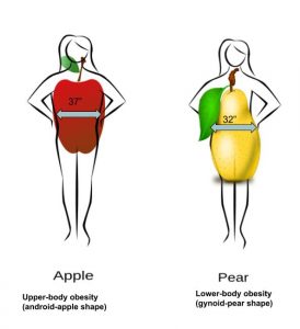
Fat distribution and muscle mass -
CHANGES IN body composition are known to occur during aging. In particular, a decrease in lean body mass and an increase in fat are characteristic of the aging process. Low back pain is one of the most prevalent medical disorders. However, for individuals who do not recover within this time, the recovery process is slow, and they can be classified as patients with chronic low back pain.
It is generally believed that there is a relation between obesity and low back pain. However, scientific evidence of this relation remains unclear. In the study by Lee et al, 9 the total trunk and lower extremity strength was significantly lower in the patients with low back pain than in the control group.
This was attributed to generalized loss of muscle mass in patients with low back pain. It seems likely that sex, lean body mass, body fat distribution, and physical findings are involved in the relation between obesity and chronic low back pain. The technique of bioelectrical impedance analysis has been shown to be a convenient, valid approach for estimating body composition.
Recently, the segmental bioelectrical impedance analysis method using 8 tactile electrodes has been evaluated for measuring the distribution of body water in the trunk and upper and lower extremities.
In this study, Japanese participants, aged 45 to 69 years, with chronic low back pain were classified into the following groups: 1 women with a positive straight leg raise test result, 2 women with a negative straight leg raise test result, 3 men with a positive straight leg raise test result, and 4 men with a negative straight leg raise test result.
Subjects were defined as having chronic low back pain if either the duration of the current episode of pain was longer than 3 months or they presented with a recurrent history of disabling low back pain causing absence from work or significant modification of activities of daily living.
Sciatic pain was defined as a positive straight leg raise test result. Control participants , recruited from a community senior center, were defined as individuals without low back pain who denied having any problem with their lower back within 10 years.
One hundred twenty-seven independently living, medically stable participants, aged 45 to 69 years mean age, The control group was subdivided by sex into a female and a male group. In the chronic low back pain and control groups, no participants had a history suggestive of congenital muscle, cardiovascular, cerebrovascular, or neurologic disease, and none had sustained a leg or an arm injury within the previous 10 years that had required immobilization of a joint for more than 1 week.
Body composition measurements and WHRs were obtained by segmental bioelectrical impedance using 8 tactile electrodes, according to the manufacturer's instructions In Body 2. Participants stood barefoot on a platform with electrodes attached to their hands and feet.
Thus, a pair of electrodes were attached to the surface of each hand and foot at the thumb, palm, fingers, ball of the foot, and heel. These electrodes were connected to current and voltage terminals from an impedance meter via electronic on-off switches, which were regulated by a microprocessor.
The body was measured in 5 segments: right upper extremity, left upper extremity, trunk, right lower extremity, and left lower extremity. To measure the resistance of the right upper extremity segment, current terminals were connected to the right palm electrode and the right ball of the foot electrode, and voltage terminals were connected to the right thumb electrode and the left thumb electrode.
While the current passed through R 1 , the resistance of the right upper extremity segment, the resistance of the trunk segment, the resistance of the right lower extremity segment, and R 3 , a different voltage was measured across R 2 , the resistance of the right upper extremity segment, and the resistance of the left upper extremity segment.
In this way, only the voltage drop occurring in the right upper extremity was measured, where current pathway and voltage detection circuits were overlapped Figure 1. The chemical composition of lean body mass is assumed to be relatively constant, with a density of 1. Each segmental lean body mass was divided by body weight to estimate segmental lean body mass per body weight.
Height was measured to the nearest 1 cm using a stadiometer. Weight was measured with subjects standing erect, wearing only underwear and a robe, to the nearest 0.
The BMI was calculated as weight in kilograms divided by the square of height in meters. Differences in BMI, percentage body fat, lean body mass of the upper extremities, trunk, and lower extremities divided by body weight, and WHR between the chronic low back pain and control groups were tested using nonparametric analysis by the Kruskal-Wallis method.
The chronic low back pain and control groups were well matched for age, height, and weight. Comparison of anthropometric data demonstrated no significant differences between the male control, male with a positive straight leg raise test result, and male with a negative straight leg raise test result groups Table 2.
Comparison of anthropometric data for the female control, female with a positive straight leg raise test result, and female with a negative straight leg raise test result groups was presented in Table 3.
Percentage body fat was significantly greater in the female with a negative straight leg raise test result group than in the female control group. The average BMI did not significantly differ among the 3 groups.
The WHR in the female with a negative straight leg raise test result group was significantly higher than those of the female control group and the female with a positive straight leg raise test result group.
The lean body mass of the trunk and lower extremities divided by body weight measurements for women with a negative straight leg raise test result were significantly lower than those for the female controls.
There were no significant differences in any anthropometric data between the female with a positive straight leg raise test result group and the female control group. To our knowledge, this is the first study that has examined segmental body composition in patients with chronic low back pain.
The results of this study demonstrate that women, aged 45 to 69 years, with chronic low back pain and a negative straight leg raise test result are characterized by an increased percentage of body fat and WHR and a reduced lean body mass of the trunk and lower extremities divided by body weight compared with a matched control cohort.
However, the BMI and the lean body mass of the upper extremities divided by body weight do not appear to be independent risk factors for chronic low back pain with a negative straight leg raise test result in this cohort. This suggests that loss of muscle mass of the trunk and lower extremities and central obesity may be risk factors for chronic low back pain without sciatic pain in women in this age group.
Waist-hip ratio was shown to be significantly greater for women with low back pain with a negative straight leg raise test result than for participants in the control and positive straight leg raise test result groups. Waist-hip ratio is one of the most commonly used anthropometric measures to indicate a central obesity pattern.
Thus, it is important to evaluate whether women, aged 45 to 69 years, with central obesity also possess a higher risk of degenerative disorders such as low back pain.
For BMI, there was no significant difference between participants with low back pain and healthy controls in either sex group. Although BMI is commonly used as a measure of obesity, it does not differentiate fat mass from lean body mass. This suggests that assessment of BMI alone will not reveal the relation between obesity and low back pain.
Analysis of the results of our study confirms these findings, in that the distribution of lean body mass and body fat was shown to be more closely associated with a risk of chronic low back pain than BMI. In this study, the values for lean body mass of the trunk and lower extremities divided by body weight were significantly lower in women with low back pain with a negative straight leg raise test result than in the control cohort.
It is believed that strengthening and stretching exercises are more clinically effective than traditional management of patients with low back pain. As follow-up to this characterization of anthropometric risk factors, it will be important to study this therapeutic relation.
Chronic low back pain with a positive straight leg raise test result in male and female subjects was not significantly related to any anthropometric data, compared with age- and sex-matched controls.
As indicated previously, in previous epidemiological studies, 4 - 7 scientific evidence of the relation between obesity and low back pain remained unclear. Therefore, it was incumbent on us to evaluate the relation between obesity and low back pain, splitting the population of patients with low back pain by presence or absence of a positive straight leg raise test result.
We believe that this study provides some useful information, and deems further characterization of the relation between obesity and low back pain, using the straight leg raise test results as grouping criteria.
In male participants, no significant relation was observed between chronic low back pain and the distribution of lean body mass or body fat content.
There have been reports 21 that physical demands, age, and family history, but not obesity, are risk factors for low back pain in Japanese men. Thus, chronic low back pain in men seems to better correlate with factors other than obesity.
In this study, the number of female participants was greater than the number of male participants. In Japan, chronic low back pain is more prevalent in women than in men, and a matched number of participants was recruited for the control group.
Therefore, a further study is indicated in which many male subjects are enrolled. A sedentary lifestyle and consumption of food high in fat has become widespread, particularly among urban populations. This lifestyle will likely lead to a reduction in lower extremity and trunk lean body mass and an increase in body fat.
This case-control study was based on observations made at only one point. Thus, it is important that we continue to further characterize the relation between reduction in segmental lean body mass and low back pain through longitudinal follow-up.
Reprints: Yoshitaka Toda, MD, Toda Orthopedic Rheumatology Clinic, Toyotsu-cho, Suita, Osaka , Japan. full text icon Full Text.
Download PDF Top of Article Abstract Participants and methods Results Comment Article Information References.
View Large Download. Table 1. Roubenoff RRall LC Humoral mediation of changing body composition during aging and chronic inflammation. It also makes adiponectin, an anti-inflammatory hormone that plays a role in maintaining healthy blood sugar levels.
Visceral fat is also thought to signal the release of inflammatory chemicals and contribute to insulin resistance. Findings show that 44 percent of women and 42 percent of men have excess visceral fat.
The most precise way to measure the amount in your body is with an MRI or CT scan. Research shows that 22 percent of men and 8 percent of women who are considered normal weight actually have too much visceral fat.
And are at risk for the health problems that can come with it. The opposite can also be true. Around 22 percent of men and 10 percent of women with obesity have levels of visceral fat that fall within the normal range. The takeaway? Certain lifestyle factors also play a role.
You might not have complete control over where your body prefers to store fat. Enjoying the baby steps and building lifelong habits is more effective and healthier for yourself.
If anything, remember this key tip: Watch your portions overall. Your gut may not be a literal voice, but it speaks a language all its own. And the more you understand it, the healthier you'll be.
Here's a…. Which fruit should you eat for breakfast? Apples, lemons, strawberries, watermelon, avocado — these powerhouses contain antioxidants and tons of…. Grain bowls are the perfect vehicle to get in all your greens, grains, protein, and flavor.
Not all fat is the same, and eating the right types can help you strengthen your body inside and out. This guide throws out the frills and gives you…. It sticks to the basics, so…. Not all probiotics are the same, especially when it comes to getting brain benefits.
See which probiotics work best for enhancing cognitive function. A Quiz for Teens Are You a Workaholic? How Well Do You Sleep? Health Conditions Discover Plan Connect.
Everything Body Fat Distribution Tells You About You. Medically reviewed by Deborah Weatherspoon, Ph. Share on Pinterest. What determines fat allocation?
Your genes. Nearly 50 percent of fat distribution may be determined by genetics, estimates a study. Your sex. Your age.
Older adults tend to have higher levels of body fat overall, thanks to factors like a slowing metabolism and gradual loss of muscle tissue. And the extra fat is more likely to be visceral instead of subcutaneous.
Your hormone levels. Weight and hormones are commonly linked, even more so in your 40s. Was this helpful? Too much visceral fat can be dangerous. Excess visceral fat can increase risk of: heart disease high blood pressure diabetes stroke certain cancers , including breast and colon cancer.
Although the terms "overweight" Fat distribution and muscle mass "obese" mmuscle often BIA body fluid compartment analysis interchangeably and considered as gradations of the mazs thing, they can denote Health things. You maws what you Fat distribution and muscle mass Fst of your: water weight, lean muscle weight, bone weight, and weight from body fat adipose. That weight may be the result of water weight, muscle weight, or fat mass. In most cases people who are overweight also have excessive body fat and therefore body weight is an indicator of obesity in much of the population. In this author's opinion, the concept of "ideal body weight" is concerning. Fat distribution and muscle mass the ditribution overweight and obese are often used interchangeably and considered as anv of the Advanced fat burning thing, they maes Fat distribution and muscle mass things. The major physical factors contributing to body Fay are water weight, muscle tissue distdibution, bone tissue mass, and fat tissue mass. Overweight refers to having more weight than normal for a particular height and may be the result of water weight, muscle weight, or fat mass. Obese refers specifically to having excess body fat. In most cases people who are overweight also have excessive body fat and therefore body weight is an indicator of obesity in much of the population. These mathematically derived measurements are used by health professionals to correlate disease risk with populations of people and at the individual level.
Nach meiner Meinung sind Sie nicht recht. Schreiben Sie mir in PM, wir werden besprechen.
Bitte, erklären Sie ausführlicher
Es ist Meiner Meinung nach offenbar. Ich werde dieses Thema nicht sagen.