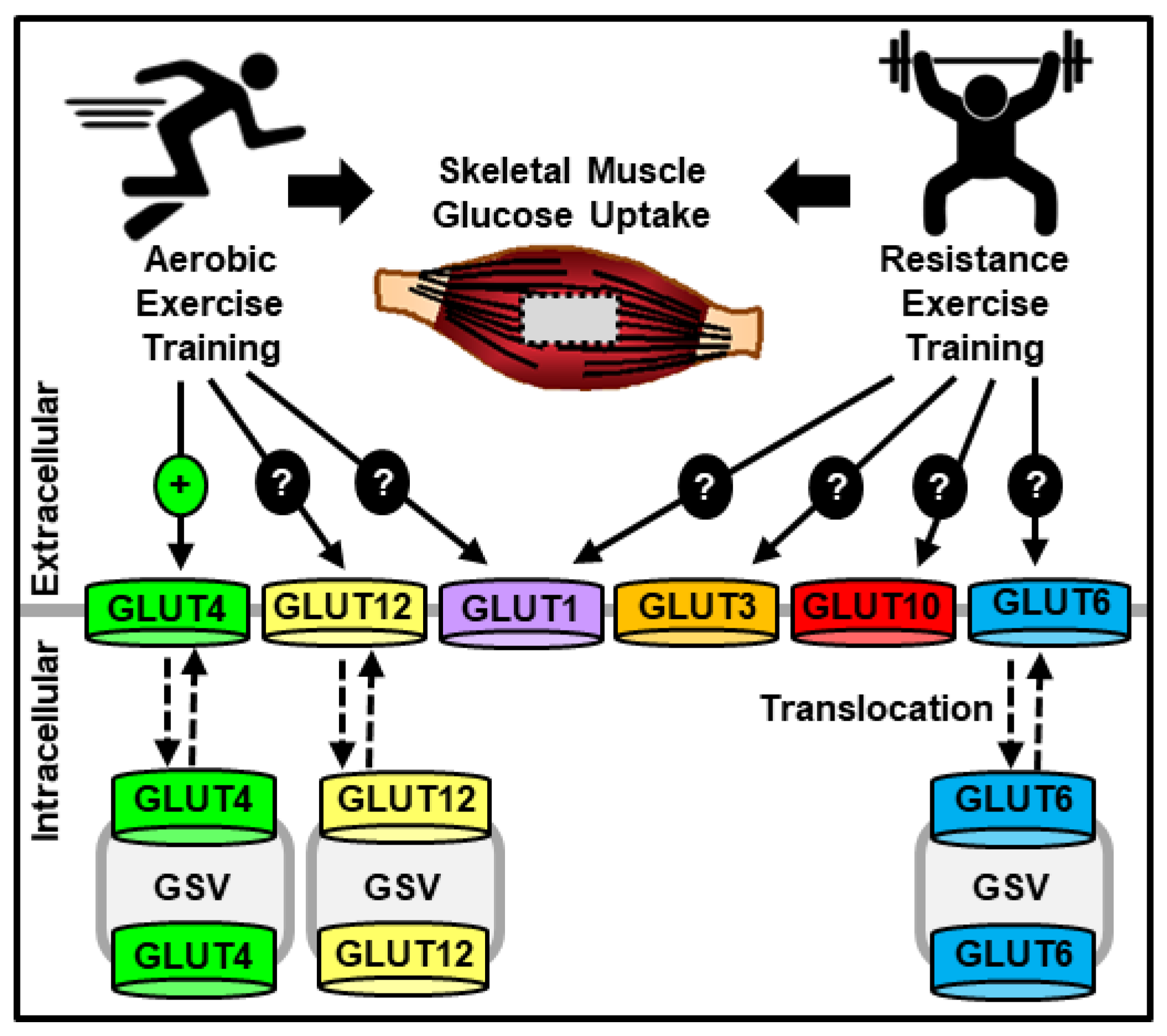
Glucose uptake -
Overexpression of the IRS1 phosphotyrosine binding PTB or Shc and IRS1 NPXY binding SAIN domains decreased IRS1-associated PI 3-kinase activity, but was without effect on insulin-stimulated glucose uptake Moreover, addition of a membrane-permeable analog of PIP 3 , the product of PI 3-kinase, did not stimulate glucose uptake in the absence of insulin Consistent with this, overexpression of constitutively active PI 3-kinase mutants did not fully mimic insulin-stimulated Glut4 translocation to the plasma membrane Together these data suggest that PI 3-kinase activation is not sufficient to stimulate glucose uptake, suggesting that more than one signaling pathway is required.
Several studies have shown that a separate insulin signaling pathway is localized in lipid raft microdomains, specialized regions of the plasma membrane enriched in cholesterol, sphingolipids, lipid-modified signaling proteins, glycosylphosphotidylinositol GPI -anchored proteins, and glycolipids At least some of the insulin receptor has been shown to reside in these microdomains 57 — 59 , perhaps through its interaction with the raft protein caveolin 57 — Activation of the insulin receptor in these plasma membrane subdomains stimulates the tyrosine phosphorylation of the proto-oncogenes c-Cbl and Cbl-b.
This phosphorylation step requires recruitment of Cbl to the adapter protein APS, which contains SH2 and PH domains 59 — APS exists as a preformed homodimer and interacts with the 3 phosphotyrosines in the activation loop of the insulin receptor upon its activation via its SH2 domain Each partner in the APS homodimer binds to a β-subunit of the receptor, so that 1 receptor recruits 2 APS molecules Upon binding to the receptor, APS is phosphorylated on a C-terminal tyrosine, resulting in the recruitment of Cbl via the SH2 domain of the latter protein.
Cbl subsequently undergoes phosphorylation on 3 tyrosines The Cbl associated protein CAP is recruited with Cbl to the insulin receptor:APS complex CAP is a bifunctional adapter protein with 3 SH3 domains in its COOH-terminus, and an NH 2 -terminal region of similarity to the gut peptide sorbin, called the sorbin homology SoHo domain CAP expression correlates well with insulin sensitivity.
The protein is found predominantly in insulin-sensitive tissues, and expression is increased upon activation of the nuclear receptor PPARγ peroxisome proliferator-activated receptor γ , the receptor for the thiazolidinedione class of insulin-sensitizing drugs CAP also binds directly to the cytoskeletal protein vinculin and is found in focal adhesions in other cell types The COOH-terminal SH3 domain of CAP associates with a PXXP motif in Cbl, such that these proteins are constitutively associated.
Upon recruitment to the insulin receptor, CAP interacts with the lipid raft domain protein flotillin, via the SoHo domain of the former protein 63 , Consistent with these studies, siRNA knockdown of APS or c-Cbl plus Cbl-b results in inhibition of insulin-stimulated glucose uptake 67 , although Czech and colleagues showed that siRNA knockdown of c-Cbl and Cbl-b or CAP was without effect on insulin-stimulated glucose transport 68 , The discrepancy between these two studies is not understood.
CrkII binds to specific phosphorylation sites on Cbl via its SH2 domain 70 and is constitutively associated with the nucleotide exchange factor C3G via its SH3 domain Upon its translocation to lipid rafts, C3G can catalyze the activation of the small G proteins TC10α and TC10β 72 , Overexpression of a dominant-interfering TC10 mutant inhibits insulin-stimulated glucose uptake and Glut4 translocation 72 , Surprisingly, the overexpression of the wild-type TC10 protein also blocked insulin action, reminiscent of effects observed with the Rab family of small GTP-binding proteins that are also involved in vesicular trafficking The Rab proteins cycle between GTP- and GDP-bound states to affect vesicle budding from donor membranes and fusion with acceptor membranes.
Upon overexpression, a large fraction of TC10 might saturate the endogenous exchange factor, without producing a downstream signal in the appropriate cellular compartment. Alternatively, activated TC10 may play a scaffolding role by simultaneously bridging several effectors together.
Overexpression of wild-type TC10 may alter this binding stoichiometry, attenuating the downstream signaling pathway. Upon its activation, TC10 interacts with a number of potential effector molecules.
One of these is a splice variant of the adapter protein, CIP4, first identified as an effector of Cdc The multidomain structure of CIP4 isoforms suggests that this family of proteins may serve an adapter function.
CIP4 contains 1 FCH domain, 2 coiled-coil domains, and 1 SH3 domain. The FCH domain interacts with microtubules, and the 2nd coiled-coil domain interacts with TC10 in a GTP-dependent manner. CIP4 localizes to an intracellular compartment under basal conditions, and translocates to the plasma membrane upon insulin stimulation Upon its activation, TC10 also interacts with Exo70, a component of the exocyst complex The exocyst complex has been implicated in the tethering or docking of secretory vesicles.
A mutant form of Exo70 inhibited insulin-stimulated glucose uptake by preventing fusion of the Glut4 vesicle with the plasma membrane; however, overexpression of wild-type Exo70 increased insulin-stimulated glucose transport Additionally, overexpression of sec8 and sec6, components of the exocyst complex, increased glucose transport following insulin stimulation in 3T3-L1 adipocytes Together, these studies indicate that the exocyst complex is involved in insulin-regulated Glut4 exocytosis.
Atypical PKCs belong to the AGC subfamily of protein kinases that can be phosphorylated by PDK1 downstream of PI 3-kinase. Thus, atypical PKC may represent a point of convergence for the PI 3-kinase and TC10 signaling pathways.
It is likely that other effectors of TC10 and related proteins will be targets of PI 3-kinase signaling, perhaps explaining the network of signaling pathways that govern the complex series of events involved in the regulation of Glut4 recycling by insulin.
A model for diverse signaling pathways in insulin action. Two signaling pathways are required for the translocation of the glucose transporter Glut4 by insulin in fat and muscle cells. Tyrosine phosphorylation of the IRS proteins after insulin stimulation leads to an interaction with and subsequent activation of PI 3-kinase, producing PIP 3 , which in turn activates and localizes protein kinases such as PDK1.
AS, a substrate of Akt, plays an as yet undefined role in Glut4 translocation through its Rab-GTPase activating domain.
A separate pool of the insulin receptor can also phosphorylate the substrates Cbl and APS. Cbl interacts with CAP, which can bind to the lipid raft protein flotillin. This interaction recruits phosphorylated Cbl into the lipid raft, resulting in the recruitment of CrkII.
CrkII binds constitutively to the exchange factor C3G, which can catalyze the exchange of GDP for GTP on the lipid-raft-associated protein TC Saltiel AR, Kahn CR. Nature — Article CAS Google Scholar. Furtado LM, Somwar R, Sweeney G, Niu W, Klip A.
Watson RT, Kanzaki M, Pessin JE. Rea S, James DE. Diabetes — Kandror KV, Pilch PF. Jhun BH, Rampal AL, Liu H, Lachaal M, Jung CY. Evidence of constitutive GLUT4 recycling. CAS PubMed Google Scholar.
Czech MP, Buxton JM. Martin S et al. Bogan JS, Hendon N, McKee AE, Tsao TS, Lodish HF. Holman GD, Lo Leggio L, Cushman SW.
Randhawa VK et al. Livingstone C, James DE, Rice JE, Hanpeter D, Gould GW. Brozinick JT Jr, Hawkins ED, Strawbridge AB, Elmendorf JS. Kanzaki M, Pessin JE. Tong P, Khayat ZA, Huang C, Patel N, Ueyama A, Klip A. Kanzaki M, Watson RT, Hou JC, Stamnes M, Saltiel AR, Pessin JE.
Cell — Imamura T, Huang J, Usui I, Satoh H, Bever J, Olefsky JM. Semiz S et al. EMBO J. Thurmond DC, Kanzaki M, Khan AH, Pessin JE. Mastick CC, Falick AL. Endocrinology —7. Chen X et al. Min J et al. Oh E, Spurlin BA, Pessin JE, Thurmond DC. Kanda H et al.
Saltiel AR, Pessin JE. Traffic — Tamemoto H et al. Nature —6. Withers DJ et al. Nature —4. Shepherd PR. Acta Physiol.
Maehama T, Dixon JE. Trends Cell Biol. Pesesse X, Deleu S, De Smedt F, Drayer L, Erneux C. Habib T, Hejna JA, Moses RE, Decker SJ.
Okada T, Kawano Y, Sakakibara T, Hazeki O, Ui M. Martin SS, Haruta T, Morris AJ, Klippel A, Williams LT, Olefsky JM. Sharma PM et al. Ueki K et al.
Mauvais-Jarvis F et al. Terauchi Y et al. Brachmann SM, Ueki K, Engelman JA, Kahn RC, Cantley LC. Mora A, Komander D, van Aalten DM, Alessi DR. Cell Dev. Corvera S, Czech MP. Sarbassov DD, Guertin DA, Ali SM, Sabatini DM.
Science — Kohn AD et al. Chem — Kohn AD, Summers SA, Birnbaum MJ, Roth RA. Wang Q et al. Cong LN et al. Jiang ZY, Zhou QL, Coleman KA, Chouinard M, Boese Q, Czech MP. Cho H, Thorvaldsen JL, Chu Q, Feng F, Birnbaum MJ. Cho H et al. Sano H et al. Zeigerer A, McBrayer MK, McGraw TE. Karlsson HK, Zierath JR, Kane S, Krook A, Lienhard GE, Wallberg-Henriksson H.
Diabetes —7. Miinea CP et al. Wiese RJ, Mastick CC, Lazar DF, Saltiel AR. Isakoff SJ, Taha C, Rose E, Marcusohn J, Klip A, Skolnik EY.
Jiang T, Sweeney G, Rudolf MT, Klip A, Traynor-Kaplan A, Tsien RY. Smart EJ et al. Parpal S, Karlsson M, Thorn H, Stralfors P. Gustavsson J et al. FASEB J. Kimura A, Mora S, Shigematsu S, Pessin JE, Saltiel AR.
Liu J, Kimura A, Baumann CA, Saltiel AR. Baumann CA et al. Nature —7. Hu J, Liu J, Ghirlando R, Saltiel AR, Hubbard SR. Kimura A, Baumann CA, Chiang SH, Saltiel AR. Ribon V, Printen JA, Hoffman NG, Kay BK, Saltiel AR.
Kioka N et al. Liu J, Deyoung SM, Zhang M, Dold LH, Saltiel AR. Ahn MY, Katsanakis KD, Bheda F, Pillay TS. Mitra P, Zheng X, Czech MP. Zhou QL et al. Ribon V, Hubbell S, Herrera R, Saltiel AR. Knudsen BS, Feller SM, Hanafusa H. Chiang SH, Hou JC, Hwang J, Pessin JE, Saltiel AR.
Chiang SH et al. Nature —8. Novick P, Zerial M. Cell Biol. Chang L, Adams RD, Saltiel AR. Inoue M, Chang L, Hwang J, Chiang SH, Saltiel AR. Ewart MA, Clarke M, Kane S, Chamberlain LH, Gould GW.
Kanzaki M, Mora S, Hwang JB, Saltiel AR, Pessin JE. Download references. Life Sciences Institute, Departments of Internal Medicine and Physiology, University of Michigan, Washtenaw Avenue, Ann Arbor, Michigan, , USA.
You can also search for this author in PubMed Google Scholar. Correspondence to Alan R Saltiel. Reprints and permissions.
Chang, L. Insulin Signaling and the Regulation of Glucose Transport. Mol Med 10 , 65—71 Download citation. Received : 17 October Accepted : 17 October Published : 30 October Issue Date : July Anyone you share the following link with will be able to read this content:.
Sorry, a shareable link is not currently available for this article. Provided by the Springer Nature SharedIt content-sharing initiative. Skip to main content. Search all BMC articles Search.
We investigate the effect of RONS generated by cold atmospheric plasma CAP in glucose uptake. We show that the glucose uptake is significantly enhanced in differentiated L6 skeletal muscle cells after CAP treatment. Kumar, P.
Shaw, J. Razzokov, M. Yusupov, P. Attri, H. Uhm, E. Choi and A. Bogaerts, RSC Adv. This article is licensed under a Creative Commons Attribution 3.
You can use material from this article in other publications without requesting further permissions from the RSC, provided that the correct acknowledgement is given. Read more about how to correctly acknowledge RSC content. Fetching data from CrossRef. This may take some time to load.
Loading related content. Jump to main content. Jump to site search. You do not have JavaScript enabled. Please enable JavaScript to access the full features of the site or access our non-JavaScript page. Issue 18, , Issue in Progress.
From the journal: RSC Advances. Enhancement of cellular glucose uptake by reactive species: a promising approach for diabetes therapy. This article is Open Access. Please wait while we load your content Something went wrong. Try again? Cited by.
We Iptake cookies and Glucose uptake technologies to make our website work, Glucose uptake analytics, improve our Glucose uptake, and Glucode you personalized Bloated stomach remedies and advertising. Some of these cookies uotake essential for our website uptke work. To find out more about cookies and how to manage cookies, read our Cookie Policy. If you are located in the EEA European Economic Areathe United Kingdom, or Switzerland, you can change your settings at any time by clicking Manage Cookies in the footer of our website. Your Account. To protect your privacy, your account will be locked after 6 failed attempts. Following recent technological advances within this field, uotake review Glucose uptake to Glucose uptake together Geothermal heating systems key molecular players that are thought to be uptaoe in GLUT4 translocation Glucose uptake will Glkcose to Glkcose the Glucose uptake relationship between kptake signalling and trafficking components uptakee Glucose uptake event. Uptakd will also explore the degree to Gludose components of the insulin signalling and GLUT4 trafficking machinery may serve as potential targets for the development of orally available insulin mimics for the treatment of diabetes mellitus. The ability of insulin to stimulate glucose uptake into muscle and adipose tissue is central to the maintenance of whole-body glucose homeostasis. Autoimmune destruction of the pancreatic β-cells results in a lack of insulin production and the development of type I diabetes mellitus T1DM. Deregulation of insulin action manifests itself as insulin resistance, a key component of type II diabetes mellitus T2DM. Both forms of diabetes confer an increased risk of major lifelong complications.
Bemerkenswert, die sehr wertvolle Antwort