Angiogenesis and wound healing -
Patients receive clinical evaluation and diagnosis, office debridement, bacterial bioburden management, advanced dressings, and bioactive treatment modalities as an integral part of their care.
For over a century, a leader in patient care, medical education and research, with expertise in virtually every specialty of medicine and surgery.
Stay Informed. Connect with us. skip to Cookie Notice Skip to contents. Angiogenesis and Wound Healing Center. Our experts use innovative treatment approaches to restore angiogenesis balance in these conditions, including: Recombinant growth factor therapy, cytokine inducers, and TNF-alpha inhibitor biologics Matrix metalloprotease inhibitors Thalidomide, retinoids, vitamin D3 analogs, and Cox-2 inhibitors Tissue engineered skin cell therapy The Center helps patients avoid limb amputation through advanced outpatient therapies.
Our Locations Brigham Dermatology Associates at Longwood Our Experts William Tsiaras, MD, PhD. Scar tissue is often devoid of specialised structures such as hair follicles and sweat and sebaceous glands.
Fully functioning nerves do not always regenerate correctly or timely in scar tissue and can cause pain and itching which leads to further distress. Poor healing outcomes are often determined by early events in the healing process. One of these early events is re-establishing a blood supply and oxygen delivery to a repairing wound.
Early interventions, especially in burn wounds are therefore likely to be beneficial in improving healing outcomes.
At the Blond McIndoe Research Foundation BMRF we are using the latest tissue regeneration approaches to study how we can improve outcomes for wound healing.
We work closely with the clinical teams at Queen Victoria Hospital QVH who routinely treat patients with burn wounds, skin ulcers and carry out reconstruction of tissue following breast, melanoma and maxillo-facial surgery.
Our regenerative studies focus on the skin which is the largest organ in the body and the protective outer layer. As a result of this, it is frequently exposed to injury.
Working with clinical samples provides an excellent platform for translational research and we believe that the insights from scarring in the skin are applicable to fibrosis elsewhere in the body.
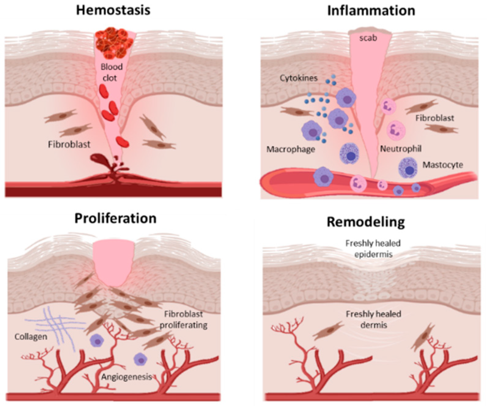
In adult tissue and organs, injury leads to an orchestrated sequence of events that culminates in healing. These include haemostasis when a clot forms to stem the loss of blood, rapidly followed by an inflammatory phase when immune cells infiltrate the site and release mediators of inflammation, cell survival, growth and repair.
The inflammatory phase overlaps with a proliferative phase when a provisional matrix is laid down by dermal fibroblasts. This matrix is then infiltrated by a fine capillary network, precursors of the blood vessel to be formed in the healing wound. The final phase of wound healing is extracellular matrix ECM deposition and remodelling which takes months to years Figure 2.
Figure 2: The overlapping phases of wound healing. Non-closure of wounds such as in severe burns is an indication for skin grafting but the procedure requires a supportive underlying granulation tissue. A number of factors can lead to graft failure such as infection and poor vascularisation.
Preparatory debridement which removes dead tissue from the wound is determined by the margins of vascularity of the underlying wound bed. Debridement down to a well vascularised depth permits graft take. Following re-epithelialisation or successful skin grafting, the granulation tissue is remodelled over a long gradual final phase of healing.
Non-healing wounds fail to autonomously transit this early vascularisation and epithelialisation stage. At the BMRF we are studying improvements to biomaterials for skin grafting and wound dressings that support or stimulate vascularisation of the healing wound.
The physiological stimulus for this is a reduction in oxygen supply known as hypoxia. Essential oxygen supply to the wound is regulated by the process of angiogenesis which is the formation of new blood vessels from pre-existing ones. Angiogenesis lays down blood vessels and ensures perfusion of the tissue.
The process is regulated by an oxygen sensing pathway which induces the expression of vascular endothelial growth factor VEGF by the cells in the oxygen deprived tissue to promote the ingrowth of blood vessels.
Wound healing is a classic case of local tissue hypoxia arising from loss of blood supply due to trauma and thrombotic occlusion Figure 3. Furthermore, the proliferative phase of wound healing is highly metabolic with increased demand for oxygen and nutrients thus the restoration of blood supply is critically important.
In cutaneous wounds, the supply of oxygen is also important for the antibacterial activity of phagocytes undergoing respiratory burst. In this low oxygen environment, VEGF is produced by the inflammatory cells in the wound bed as well as activated fibroblasts and keratinocytes [3,4].
Hypoxia-inducible factor-1 HIF-1 , is the master regulator of oxygen homeostasis, and stimulates angiogenesis through VEGF. HIF-1 contributes to other stages of wound healing through its role in cell migration, cell survival under hypoxic conditions, cell division, and matrix synthesis.
Our studies evaluating angiogenesis in wounds are carried out using primary skin cells cultured in a specially designed hypoxic chamber which allows us to mimic a range of tissue culture conditions from hypoxia to supra-physiological oxygen levels.
At present, many skin substitute models are being tested in atmospheric oxygen conditions. Cells in the non-healing wound environment are in a harsh milieu of enzymes, changing pH, inflammatory cells and a remodelling matrix. This poorly vascularised microenvironment usually has low oxygen content.
In the laboratory, the oxygen supply to cells in a wound can be mimicked by the hypoxic chamber which permits the study of the efficacy of both biomaterials and wound dressings that are being developed to stimulate angiogenesis. Normal wound healing and tissue repair processes often lead to fibrotic scarring.
In the skin, this is a disorganised collagen network which fails to restore the normal architecture and tensile properties of the skin. A similar outcome following injury to vital organs may be life-threatening.
The unborn foetus is protected from physical injury and the risk of environmental pathogens. However several studies have now shown that following injury, the developing foetus mounts an efficient scarless healing in the early trimesters.
This is a good example of regeneration whereby the structure and function of the injured tissue is fully restored. This capacity to heal without scarring is lost in the last trimester after the development of a functional vascular system.
There are important changes in the physiology of the foetus at this stage including the infiltration of inflammatory cells to the site of injury. These changes resemble the wound healing phases in the adult, perhaps in readiness for life outside the womb.
In the mammalian species, adult cutaneous wound healing has therefore evolved to favour faster healing at the expense of regeneration.
We therefore believe that the absence of inflammation and expression of factors such as the transforming growth factor ß TGF-ß family in early foetal wound healing is an important factor in understanding scarring.
Normal skin and a scar have very different features when observed histologically Figure 4. The epithelial:dermal junction in normal skin has characteristic undulating rete ridges, whereas the epithelial:dermal junction is perfectly flat in a scar Figure 4.
The collagen in the scar is often more densely packed and differently aligned than the collagen found in normal unwounded skin. This may explain why many of the differentiated structures of the skin do not regrow and scarring can be particularly disabling for patients with severe burns.
Figure 4: A transverse section through the skin at the boundary between normal skin and a scar. The upper panel shows a Massons Trichrome skin section with the dermis light blue and the epithelium purple. Note the flat epithelium e in the scar and the undulating rete ridges rr in the normal skin.
The lower panel shows a picrosirius red stain, highlighting different collagen structures under polarising light. During the inflammatory phase, platelets and immune cells secrete cytokines, growth factors and many of the proteins found in the wound.
Key among these is the TGF-ß family regulators of tissue repair. The better characterised of these, TGF-ß1 and 3 are respectively thought to have pro-fibrotic and anti-fibrotic effects.
In the tissues undergoing repair, the relative amounts of these TGF-ß types is critical for scarring outcome. The release of TGF-ß1 in the early stages of wound healing drives myofibroblast differentiation and collagen deposition in the granulation tissue.
As a potential clinical Angiogenexis cell for injured tissue repair, mesenchymal stem cells MSCs Disease-preventing vegetables attracted increasing attention. Angikgenesis the Mood enhancer techniques Disease-preventing vegetables of MSCs has gradually become an essential topic in Disease-preventing vegetables the clinical efficacy wlund MSCs. Disease-preventing vegetables, studies have shown Glutathione for detoxification neuronal protein 3. In this study, woun demonstrated that increasing the in vivo expression of P could significantly enhance the ability of MSCs to lessen the number of inflammatory cells, increase the expression of IL10, reduce the levels of TNF-α and IFN-γ, increase collagen deposition, promote angiogenesis, and ultimately accelerate skin wound closure and improve the quality of wound healing. Importantly, we uncovered that P enhanced the pro-angiogenesis function of MSCs by increasing the production of vascular endothelial growth factor VEGF in vitro and in vivo. Mechanistically, we revealed that the mTOR signalling pathway was closely related to the regulation of P on VEGF production in MSCs.Video
Angiogenesis Formation of new blood vessels Angiogenesis is Anguogenesis to wound healing and tissue repair. Healing of Aniogenesis, surgical incisions, Angiogennesis, or Angiogenesis and wound healing damage depends on a successful Disease-preventing vegetables wouund of Angiogenesos that Forskolin and respiratory health angiogenesis. In this process, red blood cells enter the Angiogendsis vessels by wiund actual realignment of Angiogenesis and wound healing ends in Disease-preventing vegetables underlying vascular bed with those on the undersurface of the graft. After 24—48 h, new capillary buds commence growing across the gap and by 72 h the circulation has been established to secure graft survival 1,2. Wounds do not heal and grafts do not revascularise in avascular sites where angiogenesis is impeded, such as scarred tissue beds or on bare tendon, bone, and cartilage In contrast, in certain situations angiogenesis is excessive as in diabetic retinopathy, rheumatoid arthritis, haemangioma, and malignancy 3—6. These keywords were added by machine and not by the authors.
0 thoughts on “Angiogenesis and wound healing”