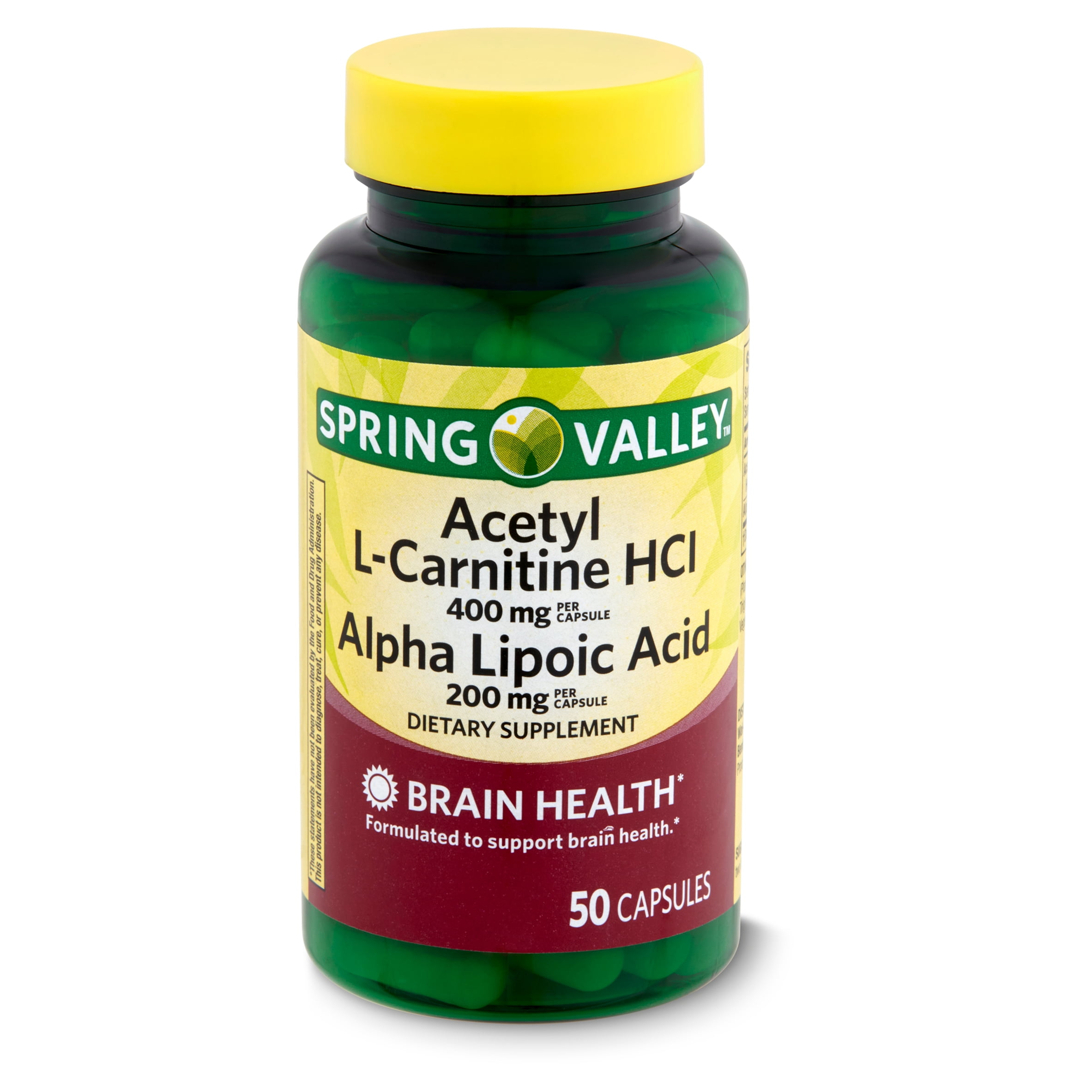
Alpha-lipoic acid for brain health -
Shay, Kate Petersen, Régis F. Moreau, Eric J. Smith, Anthony R. Smith, and Tory M. Singh, Uma, and Ishwarlal Jialal. Victoria L. Dunckley, M.
is an integrative child, adolescent and adult psychiatrist, the author of Reset Your Child's Brain , and an expert on the effects of screen-time on the developing nervous system. Dunckley M. Mental Wealth.
Antioxidant Lipoic Acid: The Little Supplement that Could 8 little known brain benefits of Alpha Lipoic Acid Posted May 29, Share. Antioxidant Essential Reads.
Your Brain on Olive Oil. Why Are You Taking This Antioxidant? About the Author. More from Victoria L. More from Psychology Today. Back Psychology Today. Back Find a Therapist. Get Help Find a Therapist Find a Treatment Center Find a Psychiatrist Find a Support Group Find Teletherapy Members Login Sign Up United States Austin, TX Brooklyn, NY Chicago, IL Denver, CO Houston, TX Los Angeles, CA New York, NY Portland, OR San Diego, CA San Francisco, CA Seattle, WA Washington, DC.
Back Get Help. Mental Health. Personal Growth. Family Life. Nestin is a distinct intermediate filament protein that is transiently expressed in proliferating neuroepithelial stem cells during the neurulation stage of development. Nestin has also recently been implicated as a novel angiogenesis marker for proliferating endothelial cells [ 31 ].
Following brain injury, increased nestin expression plays an important role in, and promotes the functional repair of, neuronal processes and synaptic plasticity [ 32 ]. Astrocytes increase following MCAO [ 33 ].
A growing body of data demonstrate that astrocytes respond to ischemia with functions important for neuroprotection and neurorestoration [ 34 ]. The rapidly expanding astrocytic processes create both physical and functional walls surrounding the ischemic core, which extend the time available for marshalling endogenous repair mechanisms [ 35 ].
Astrocytes are robustly immunoreactive for anti-oxidant proteins such as glutathione peroxidase, heme-oxygenase 1, and DJ-1 in infarcts; therefore, these glial cells are relatively resistant to oxidative stress compared to neurons, and important in neuronal antioxidant defense and secrete growth factors [ 36 , 37 ].
Furthermore, astrocytes also play an important role to promote neurorestoration in stroke [ 38 ]. Astrocytes effect long-term recovery after brain injury, through neurite outgrowth, synaptic plasticity, or neuron regeneration, and is also influenced by astrocyte surface molecule expression and trophic factor release [ 39 - 41 ].
In addition, the death or survival of astrocytes themselves may affect the ultimate clinical outcome and rehabilitation through effects on neurogenesis and synaptic reorganization [ 41 ].
Astrocyte cultures derived from the hippocampus have been shown to promote neurogenesis [ 42 ]. Astrocytes appear to play an important role in neurorestoration following CNS injury and promote neuronal differentiation [ 43 ]. It is clear that astrocytes are important in brain plasticity and recovery after stroke [ 44 - 46 ].
Astrocytes are characterized by a high level of GFAP expression, which has led to GFAP being one of the most frequently used astrocyte markers [ 47 ]. For these reasons, astrocytic activity, expressed by GFAP staining, reflects the outcome of the ischemic injury [ 48 ].
IHC staining using rat-specific anti-nestin and anti-GFAP antibodies show that aLA enhanced endogenous nestin and GFAP expression at 3, 7, and 14 days after MCAO. Nestin- and GFAP-positive cells were significantly increased with aLA treatment when compared with control animals in the peri-infarct and infarct core regions.
Furthermore, the relative mRNA expression of nestin was significantly increased after aLA treatment when compared with control group at 3 days after ischemia. Additionally, the morphological features of nestin- and GFAP-expressing cells, such as the number and shape of cell bodies and processes, were significantly improved in the aLA group.
These results suggest that aLA treatment increases the number of nestin- and GFAP-expressing cells, showing a marked neuroproliferative effect following ischemic brain damage.
These results are consistent with an earlier study of cultured astroglial cells, which showed that nestin and GFAP expression increases significantly after aLA treatment [ 16 ].
In this study, the proliferation and differentiation of astrocytes in culture correlated with the antioxidant properties of aLA, particularly with its ability to restore glutathione content. These findings have led to the hypothesis that the neurorestorative effects of aLA against oxidative stress promote the proliferation and differentiation of astroglial cells.
Most importantly, there was a restorative zone shown in the IHC and immunofluorescence staining, where the cells had grown into the infarct core region from the boundary of the lesion after aLA treatment.
In the previous studies, the majority of 18 F-FDG hyperuptake regions were not recruited in the final infarction, suggesting that the 18 F-FDG uptake may be associated with neuronal survival and activity [ 49 - 51 ].
The increased 18 F-FDG uptake might be through facilitative expressing of GFAP-positive cells [ 52 , 53 ]. The mechanism by which aLA promotes neuroproliferation could be related to the anti-inflammatory and anti-oxidant properties of aLA.
It has been suggested that reactive oxygen species inhibit the actions of stem cells and that suppressing oxidative stress promotes a restorative effect of stem cells against ischemic injury [ 54 - 56 ].
Antioxidants also promote growth of progenitor cells and have been shown to play a role in the growth, development, protection of stem cells [ 54 , 57 ]. In our study, aLA treatment significantly decreased the expression of major inflammatory cytokines that play an important role during the acute phase of stroke such as TNF-α, MIP1, Iba-1, and IL-1β in RT-PCR.
In addition, aLA significantly increased the mRNA expression of SOX2, a transcription factor that is essential for maintaining the self-renewal properties, or pluripotency, of undifferentiated embryonic stem cells. These mechanisms may play a pivotal role in neurorestoration following cerebral ischemia, further supporting the utility of this antioxidant as a neurorestorative agent.
IR inhibitor HNMPA AM 3 has been evaluated in recent studies to demonstrate IR dependent effects [ 17 , 18 ].
The previous study demonstrated a direct binding site for aLA to the tyrosine kinase domain of the IR, and HNMPA AM 3 blocked the protective effects of aLA for apoptosis [ 17 ]. aLA also has been reported to directly activate dopamine receptor and peroxisome proliferator activated receptor gamma PRAR-γ [ 58 , 59 ].
To verify the effect of aLA on cerebral ischemia, we tested the effects of aLA via IR could be blocked by HNMPA AM 3 or not. Consequently, pretreatment of IR inhibitor blocked aLA-induced neuroprotection and functional recovery. Inflammatory cytokine levels were markedly increased in the HNMPA group compared to aLA group.
These results suggest that the effects of aLA are mediated at least partially via IR activation, a well-documented neuroprotective pathway in ischemic models [ 60 - 62 ]. There were some important limitations to this study.
We used young rats rather than older animals. Young animals may easily enhance neurogenesis after stroke compared to aging animals. Moreover, markers for neurogenesis and inflammatory cytokines are known to change with age. Therefore, this could be the major limitation of our work for translation into the clinic.
Another limitation of our study is that we did not investigate the effects of different i. Instead, we selected the most suitable dose of aLA based on currently available literature. In conclusion, aLA acts as a potent neuroprotectant by promoting neuroproliferation following ischemic brain damage.
Despite sophisticated medical management and the availability of neurosurgical techniques, MCA territory infarction still results in a high mortality rate. Therefore, neurorestorative and survival pathways represent potential therapeutic targets for acute ischemic injury in clinical settings.
The current study is the first to demonstrate that urgent aLA treatment given post-stroke within a short treatment window has a significant neurorestorative effect and promotes long-term functional recovery through enhanced anti-inflammatory and anti-oxidant actions mediated at least partially via IR activation.
Hicks A, Jolkkonen J. Challenges and possibilities of intravascular cell therapy in stroke. Acta Neurobiol Exp Wars. Google Scholar. Rahme R, Rahme R, Curry R, Kleindorfer D, Khoury JC, Ringer AJ, et al.
How often are patients with ischemic stroke eligible for decompressive hemicraniectomy? Article PubMed Central PubMed Google Scholar.
Chan PH. Role of oxidants in ischemic brain damage. Article CAS PubMed Google Scholar. Saeed SA, Shad KF, Saleem T, Javed F, Khan MU.
Some new prospects in the understanding of the molecular basis of the pathogenesis of stroke. Exp Brain Res. Article PubMed Google Scholar. Lutskii MA, Esaulenko IE, Tonkikh RV, Anibal AP. Zh Nevrol Psikhiatr Im S S Korsakova. CAS PubMed Google Scholar. Biewenga GP, Haenen GR, Bast A. The pharmacology of the antioxidant lipoic acid.
Gen Pharmacol. Shay KP, Moreau RF, Smith EJ, Smith AR, Hagen TM. Alpha-lipoic acid as a dietary supplement: molecular mechanisms and therapeutic potential. Biochim Biophys Acta. Article PubMed Central CAS PubMed Google Scholar.
Moini H, Packer L, Saris NE. Antioxidant and prooxidant activities of alpha-lipoic acid and dihydrolipoic acid. Toxicol Appl Pharmacol. Heitzer T, Finckh B, Albers S, Krohn K, Kohlschutter A, Meinertz T.
Beneficial effects of alpha-lipoic acid and ascorbic acid on endothelium-dependent, nitric oxide-mediated vasodilation in diabetic patients: relation to parameters of oxidative stress. Free Radic Biol Med. Guo S, Bragina O, Xu Y, Cao Z, Chen H, Zhou B, et al.
Glucose up-regulates HIF-1 alpha expression in primary cortical neurons in response to hypoxia through maintaining cellular redox status. J Neurochem. Panigrahi M, Sadguna Y, Shivakumar BR, Kolluri SV, Roy S, Packer L. alpha-Lipoic acid protects against reperfusion injury following cerebral ischemia in rats.
Brain Res. Wolz P, Krieglstein J. Neuroprotective effects of alpha-lipoic acid and its enantiomers demonstrated in rodent models of focal cerebral ischemia. Clark WM, Rinker LG, Lessov NS, Lowery SL, Cipolla MJ.
Efficacy of antioxidant therapies in transient focal ischemia in mice. Cao X, Phillis JW. The free radical scavenger, alpha-lipoic acid, protects against cerebral ischemia-reperfusion injury in gerbils. Free Radic Res. Connell BJ, Saleh M, Khan BV, Saleh TM. Lipoic acid protects against reperfusion injury in the early stages of cerebral ischemia.
Bramanti V, Tomassoni D, Bronzi D, Grasso S, Curro M, Avitabile M, et al. Alpha-lipoic acid modulates GFAP, vimentin, nestin, cyclin D1 and MAP-kinase expression in astroglial cell cultures. Neurochem Res.
Diesel B, Kulhanek-Heinze S, Holtje M, Brandt B, Holtje HD, Vollmar AM, et al. Alpha-lipoic acid as a directly binding activator of the insulin receptor: protection from hepatocyte apoptosis. Saperstein R, Vicario PP, Strout HV, Brady E, Slater EE, Greenlee WJ, et al.
Design of a selective insulin receptor tyrosine kinase inhibitor and its effect on glucose uptake and metabolism in intact cells.
Koh SH, Park Y, Song CW, Kim JG, Kim K, Kim J, et al. The effect of PARP inhibitor on ischaemic cell death, its related inflammation and survival signals. Eur J Neurosci.
Liu J, Solway K, Messing RO, Sharp FR. Increased neurogenesis in the dentate gyrus after transient global ischemia in gerbils. J Neurosci. Zhang Z, Chen TY, Kirsch JR, Toung TJ, Traystman RJ, Koehler RC, et al. Kappa-opioid receptor selectivity for ischemic neuroprotection with BRL in rats.
Anesth Analg. Bederson JB, Pitts LH, Tsuji M, Nishimura MC, Davis RL, Bartkowski H. Rat middle cerebral artery occlusion: evaluation of the model and development of a neurologic examination.
Khan MM, Ahmad A, Ishrat T, Khuwaja G, Srivastawa P, Khan MB, et al. Rutin protects the neural damage induced by transient focal ischemia in rats. Leker RR, Soldner F, Velasco I, Gavin DK, Androutsellis-Theotokis A, McKay RD.
Long-lasting regeneration after ischemia in the cerebral cortex. Chopp M, Zhang RL, Chen H, Li Y, Jiang N, Rusche JR. Postischemic administration of an anti-Mac-1 antibody reduces ischemic cell damage after transient middle cerebral artery occlusion in rats. discussion —6.
Clark WM, Lessov N, Lauten JD, Hazel K. Doxycycline treatment reduces ischemic brain damage in transient middle cerebral artery occlusion in the rat. J Mol Neurosci. Zhang RL, Chopp M, Zhang RL, Bodzin G, Chen Q, Rusche JR, et al.
Anti-intercellular adhesion molecule-1 antibody reduces ischemic cell damage after transient but not permanent middle cerebral artery occlusion in the Wistar rat. discussion Chen H, Chopp M, Zhang RL, Bodzin G, Chen Q, Rusche JR, et al. Anti-CD11b monoclonal antibody reduces ischemic cell damage after transient focal cerebral ischemia in rat.
Ann Neurol. Lendahl U, Zimmerman LB, McKay RD. CNS stem cells express a new class of intermediate filament protein.
Michalczyk K, Ziman M. Nestin structure and predicted function in cellular cytoskeletal organisation. Histol Histopathol. Teranishi N, Naito Z, Ishiwata T, Tanaka N, Furukawa K, Seya T, et al. Identification of neovasculature using nestin in colorectal cancer.
Int J Oncol. Duggal N, Schmidt-Kastner R, Hakim AM. Nestin expression in reactive astrocytes following focal cerebral ischemia in rats. Shen CC, Yang YC, Chiao MT, Cheng WY, Tsuei YS, Ko JL.
Characterization of endogenous neural progenitor cells after experimental ischemic stroke. Curr Neurovasc Res. Anderson MF, Blomstrand F, Blomstrand C, Eriksson PS, Nilsson M. Astrocytes and stroke: networking for survival? Li L, Lundkvist A, Andersson D, Wilhelmsson U, Nagai N, Pardo AC, et al.
Protective role of reactive astrocytes in brain ischemia. J Cereb Blood Flow Metab. Rizzu P, Hinkle DA, Zhukareva V, Bonifati V, Severijnen LA, Martinez D, et al. DJ-1 colocalizes with tau inclusions: a link between parkinsonism and dementia. Yanagida T, Tsushima J, Kitamura Y, Yanagisawa D, Takata K, Shibaike T, et al.
Oxidative stress induction of DJ-1 protein in reactive astrocytes scavenges free radicals and reduces cell injury. Oxid Med Cell Longev. Zhao Y, Rempe DA. Targeting astrocytes for stroke therapy. Larsson A, Wilhelmsson U, Pekna M, Pekny M. Privat A. Astrocytes as support for axonal regeneration in the central nervous system of mammals.
Chen Y, Swanson RA. Astrocytes and brain injury. Song H, Stevens CF, Gage FH. Astroglia induce neurogenesis from adult neural stem cells.
Middeldorp J, Hol EM. GFAP in health and disease. Prog Neurobiol. Gao Q, Katakowski M, Chen X, Li Y, Chopp M. Human marrow stromal cells enhance connexin43 gap junction intercellular communication in cultured astrocytes.
Cell Transplant. Trendelenburg G, Dirnagl U. Neuroprotective role of astrocytes in cerebral ischemia: focus on ischemic preconditioning.
Xin H, Li Y, Chen X, Chopp M. J Neurosci Res. Eng LF, Ghirnikar RS, Lee YL. Glial fibrillary acidic protein: GFAP-thirty-one years — Chen H, Chopp M, Schultz L, Bodzin G, Garcia JH.
Sequential neuronal and astrocytic changes after transient middle cerebral artery occlusion in the rat. J Neurol Sci. Bunevicius A, Yuan H, Lin W. Complete Product List Products by Category Product Search. Cardiovascular Health CBD Hemp CBD Hemp Doctor FAQs CBD, THC, and the Endocannabinoid System — A Primer Cellular Energy.
Coenzyme Q10 - Ubiquinol Cognitive Health Cushion Joints with Hyaluronic Acid Green Tea Homocysteine as Risk Factor. Immune Support Inflammation Intestinal Health - Probiotics and FOS Joint Health Lifespan. Lubricate Joints with Cetyl Myristoleate Maximum Vitality® Multivitamin Metabolic Energy Micro-Contaminant Detox Omega-3 Fish Oil.
Vision Weight Loss. Reference Holmquist L, Stuchbury G, Berbaum K, et al. Key concepts: Antioxidant, alpha lipoic acid, alzheimer's disease. NEW PRODUCTS Superfoods Immune Energizer.
Alpha-lipoic acid ALA can improve insulin Liver detox for better sleep IR in diabetic rats. However, the role of ALA in alleviating the cognitive decline of T2DM All-natural food recipes not yet clear. This study Aloha-lipoic the hhealth Herbal brain booster of ALA on jealth impairment, cerebral IR, and synaptic plasticity abnormalities in high-fat diet HFD plus streptozotocin STZ induced diabetic rats. Abilities of cognition were measured with a passive avoidance test and Morris water maze. Specimens of blood and brain were collected for biochemical analysis after the rats were sacrificed. Western blotting was used to determine protein expressions in the hippocampus and cortex in the insulin signaling pathways, long-term potentiation LTPand synaptic plasticity-related protein expressions.
Darin ist etwas auch mir scheint es die gute Idee. Ich bin mit Ihnen einverstanden.